
Advanced microscopy on the timetable
This article was first published in Norwegian on our School Collaboration website.
How can we learn more about cancer cells by using advanced microscopes?
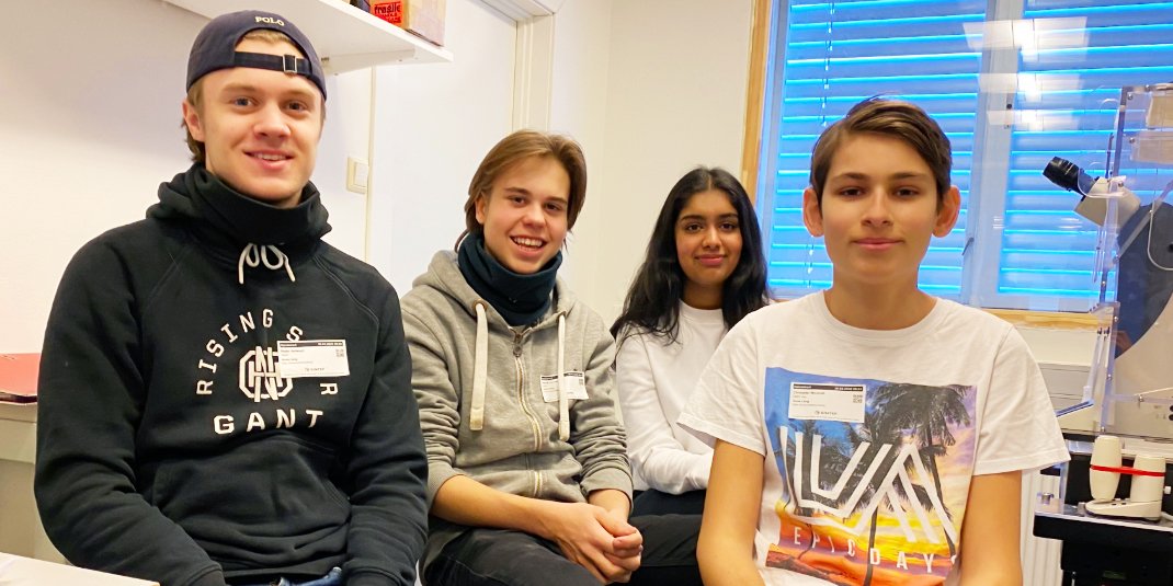
A microscope is an important tool for scientists in many different branches of research. In February, four first-year students from the Researcher programme at Ullern Upper Secondary School got to test multiple different microscopes at the Core Facility for Advanced Light Microscopy, The Gaustad node, at Rikshospitalet (Oslo University Hospital).
Isha Mohal, Peder Nerland Hellesylt, Christofer Naranjo Woxholt and Henrik Eidsaae Corneliussen are sitting in a small, rectangular room, which belongs to the research group Experimental Cancer Therapy at Oslo University Hospital.
“If you sit next to me, you can see better what I am doing,” says Emma Lång to the students.
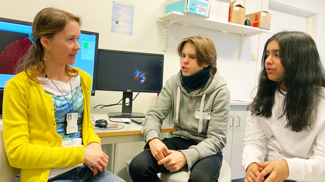
Emma Lång is a researcher in the research group Experimental Cancer Therapy. She explains to Henrik and Isha how the advanced microscope, connected to the computer behind her, can record videos of living cells. Photo: Elisabeth Kirkeng Andersen
It is the second day of the work placement for the Ullern students. Lång will show them how she is setting up a very special microscope with the somewhat cryptical name “ImageXpress Micro”.
The microscope is so special that it is the only one in the entire Oslo region and Eastern Norway. The unique thing about the microscope is that it creates videos of thousands of living cells over a long time period. This enables the researchers to understand more about how the cells move.
This is important knowledge in the research on cancer and wound healing, which this research group is working on.
The students sit down beside Lång and follow what she is doing closely. The microscope is entirely automatic, so all the settings are done on a computer. Later the same day, the students will use the microscope themselves to record videos of cells that they have been working on from the day before.
Learning from practical work
This is the first work placement for the students from the Research programme – and they are really enjoying it.
“It is fun to see what the researchers are doing and to try it out ourselves in practice,” says Peder.
“We have done some work with pipettes and worked in the laboratory at school, so we are already familiar with some of the practical handiwork. It is fun to try it out in a real research setting,” says Isha.
She likes that the placement gives some insight into what a career in research and cellular biology can be like.
“I am more interested to work in cellular biology after this placement, but I haven’t decided anything yet. I think we are learning things in an exciting way. It is practical learning and not as theoretical as it is usually in school,” says Peder.
“I absolutely see this as an opportunity to become a researcher. It is great to have so much science subjects as we have on the Researcher programme,” says Henrik and Isha agrees.
“I am very interested in the natural sciences. We have a lot of theory in school and it is fun to come out into the hospital and into companies to see how researchers work – and to try it out ourselves,” says Isha.
Christofer also thinks it is interesting, but he is more interested in data and other general subjects.
“That’s great, Christofer,” Lång says. “Research needs more people with good data knowledge. Do you see the computer over there? It costs NOK 100 000 and it will be used to develop machine learning and a technique called ‘deep learning’ on the data produced from our microscopes. Maybe in a few years time, computers will be analysing the microscope images and videos that we are recording now.”
Images of cells
Yesterday, Isha, Peder, Christofer and Henrik worked on cells in the laboratory. They learned a technique to fixate cells. Then, they coloured the cells with antibodies that turn blue when they bind to the core of the cell and with a protein called actin that turns green. Actin performs several functions in the cell, it is both inside the cell structure and functions as threads of communication between the cells.
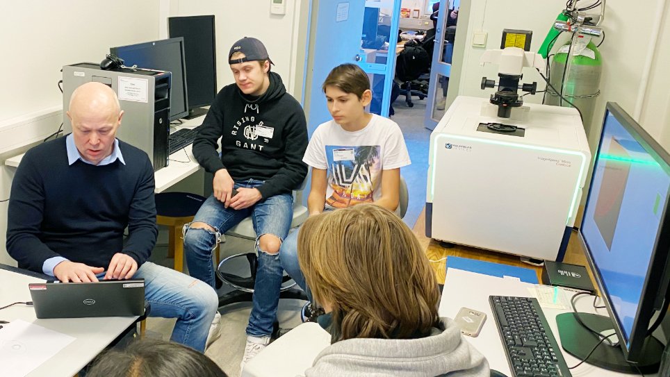
Stig Ove Bøe leads the research group that was visited by the four students from the Research programme at Ullern Upper Secondary School for two days. Here, he is preparing the images of skin cells that the students worked on the day before. Photo: Elisabeth Kirkeng Andersen
Now, the students are looking at the results uploaded to a computer in an advanced image editing software program that can visualise the cells as two- or three-dimensional.
“These are the skin cells you coloured yesterday. Can you see that the cells make up one close network? The reason for this is that it is skin and it is supposed to be impenetrable. Can you also see that the single cells act differently at the edge than closer inside? It is our job to explain why and how,” Bøe explains to the students.
The students look and nod with interest.
After the placement, researchers at Rikshospitalet (Oslo University Hospital) have worked more on the images and videos that the students created.
These have been delivered to the students and will be used when they make a presentation of the placement and everything they learned to the rest of the students at the Research programme.
You can see the cell image below.
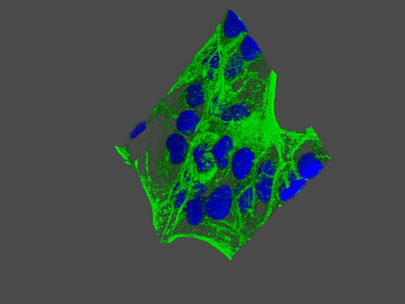
A three-dimensional image of the skin cells that the students have coloured. Photo: Emma Lång
What is cell migration?
The research group “Experimental Cancer Therapy” led by Dr Stig Ove Bøe at Rikshospitalet are researching how cells move, which is called cell migration in scientific terms.
Cell migration plays a central role in many of the body’s physiological functions, such as the immune system and wound healing. Cell migration is also essential for cancer, since cancer cells can spread from the location of the tumour to other organs of the body.
Cells use different mechanisms to migrate. They can move as single cells or they can move collectively. Thousands of cells can, for example, cooperate so they can move in the same direction.
The research group uses many different microscopy-based methods to research cell migration. They are also developing new video methods to study living cells in microscopes.
The research group is also responsibly for the daily running of the Core Facility for Advanced Light Microscopy at Oslo University Hospital. The facility gives other research groups in the Oslo region access to and guidance of the use of advanced microscopy equipment.